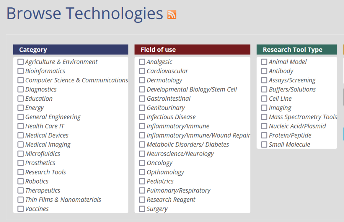
Browse Technologies

Displaying 11 - 20 of 45


Miniature Optical Coherence Tomography Probe for Real-time Monitoring of Surgery

Vanderbilt researchers have designed a forward scanning miniature intraoperative Optical Coherence Tomography (OCT) probe that can be used for diagnostic purposes and real-time monitoring of surgery within small spaces, such as endoscopic surgery, intraocular surgery, and other microsurgery.


Licensing Contact

Taylor Jordan

615.936.7505

Model-based Compression Correction Framework for Ultrasound

Vanderbilt researchers have developed a system that corrects for compressional effects in ultrasound data during soft tissue imaging. The system uses tracking and digitization information to detect the pose of the ultrasound probe during imaging, and then couples this information with a biomechanical model of the tissue to correct compressional effects during intraoperative imaging.


Licensing Contact

Philip Swaney

615.322.1067

Near-Infrared Dye with Large Stokes Shift for Simultaneous Multichannel in vivo Molecular Imaging

Fluorescent labels having near-infrared (NIR) emission wavelengths have the ability to penetrate tissue deeper than other emission wavelengths, providing enormous potential for non-invasive imaging applications. However, advancement of optical imaging (particularly NIR imaging) is hindered by the limitation of narrow Stokes shift of most infrared dyes currently available in the market. Vanderbilt researchers have developed a novel NIR dye (4-Sulfonir) for multichannel imaging that enables in vivo imaging of multiple targets due to its large Stokes shift. 4-Sulfonir with its unique large Stokes shift (~150 nm) and wide excitation spectrum could be used in parallel with other NIR dyes for imaging two molecular events simultaneously in one target.


Licensing Contact

Masood Machingal

615.343.3548
Imaging

NMR Signal Amplification by Reversible Exchange (SABRE) in Water

Vanderbilt researchers have developed a method to perform the Parahydrogen Induced Polarization (PHIP) based method of Signal Amplification by Reversible Exchange (SABRE) in aqueous media. This allows the resulting hyperpolarized molecules to be used for in vivo applications.


Licensing Contact

Chris Harris

615.343.4433
Medical Imaging

System and Methods of Using Image-guidance for Placement of Cochlear Stimulator Devices, Drug Carrier Devices, or the Like

Vanderbilt inventors have developed and tested a device (C-in) and method that would shift the current invasive, risky surgical procedure of cochlear implantation to a less invasive outpatient procedure.


Licensing Contact

Taylor Jordan

615.936.7505

Two Degrees-of-Freedom, Fluid Power Stepper Actuator Model

Vanderbilt researchers have developed a novel technology for use of a flexible fluidic actuator in MRI-guided surgical systems. This method eliminates the need for moving the patient out of the MRI machine, onto an operating table, and back in order to perform procedures. It is a safe, sterilized, and successful method to simplify MRI-guided surgical procedures.


Licensing Contact

Taylor Jordan

615.936.7505

A Method to Obtain Uniform Radio Frequency Fields in the Body for High Field MRI

Researchers at Vanderbilt have created a new approach to produce uniform radio frequency (RF) fields in the body during high field magnetic resonance imaging (MRI). Existing high field MRI machines create non-uniform RF fields that lead to non-uniform sensitivity in the generated images, also referred to as "hot" and "cold" spots. These local variations interfere with the tissue contrast of the images that radiologists depend upon to make accurate diagnoses. By generating uniform RF fields in the body, this technology provides the benefits of high field MRI without the non-uniform RF fields.


Licensing Contact

Chris Harris

615.343.4433
Medical Imaging

Enhanced Optical Imaging for the Treatment of Retinal Disease

Vanderbilt researchers have developed a system for enhancing the imaging capabilities of optical coherence tomography (OCT), a tool commonly used to monitor and treat patients with retinal disease. The image resolution of OCT, however, is intrinsically limited. Ideally, a contrast agent could be used to highlight specific parts of the retina within the image, but dye alone is largely ineffective because of the way OCT generates the image. Photothermal heating solves this problem by creating local zones of tissue expansion which can be distinctly detected by OCT. Photothermal-OCT is safe, effective, and enhances the imaging power of a tool widely used by opticians.


Licensing Contact

Philip Swaney

615.322.1067
Medical Imaging

Systems and Methods for Non-destructive Evaluation of Optical Material Properties and Surfaces

A team of Vanderbilt researchers has developed a novel system and method for non-destructive characterization of compound lenses. The approach uses optical coherence tomography and reflectance confocal microscopy to fully characterize lens geometry and glass materials, enabling accurate modeling of compound lenses.


Licensing Contact

Philip Swaney

615.322.1067
Medical Imaging

PIQASO: A rigid phantom for comprehensive end-to-end evaluation of online adaptive radiotherapy systems

There is currently no radiotherapy phantom capable of quantitatively assessing all components of an online adaptive radiotherapy (online ART) system in a comprehensive end-to-end test.Represented here is a novel, rigid phantom that can simultaneously evaluate an online ART system's image acquisition, deformable image registration, contour propagation, plan re-optimization, dose calculation, and beam delivery in a single process that is robust, quantitative, and convenient.


Licensing Contact

Masood Machingal

615.343.3548