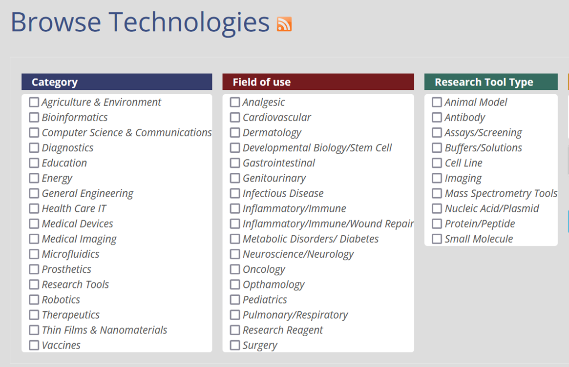
Browse Technologies

Displaying 1 - 10 of 41


Non-Invasive Skin Cancer Detection using Raman Spectroscopy-OCT System (Portfolio)

Vanderbilt University researchers have designed a system for non-invasive discrimination between normal and cancerous skin lesions. The system combines the depth-resolving capabilities of OCT technique with Raman Spectroscopy's specificity of molecular chemistry. By linking both imagining techniques into a single detector arm, the complexity, cost, and size of previously reported RS-OCT instruments have been significantly improved. The combined instrument is capable of acquiring data sets that allow for more thorough assessment of a sample than existing optical techniques.


Licensing Contact

Ashok Choudhury

615.322.2503

Systems and Methods for Optical Stimulation of Neural Tissues (Portfolio)

Vanderbilt researchers have developed a novel technique for contactless simulation of the central nervous system.  This involves the use of infrared neural stimulation (INS) to evoke the observable action potentials from neurons of the central nervous system.  While infrared neural stimulation of the peripheral nervous system was accomplished almost a decade ago, this is the first technique for infrared stimulation of the central nervous system. This technology has been protected by a portfolio of issued patents.


Licensing Contact

Ashok Choudhury

615.322.2503

Near-Infrared Dye with Large Stokes Shift for Simultaneous Multichannel in vivo Molecular Imaging

Fluorescent labels having near-infrared (NIR) emission wavelengths have the ability to penetrate tissue deeper than other emission wavelengths, providing enormous potential for non-invasive imaging applications. However, advancement of optical imaging (particularly NIR imaging) is hindered by the limitation of narrow Stokes shift of most infrared dyes currently available in the market. Vanderbilt researchers have developed a novel NIR dye (4-Sulfonir) for multichannel imaging that enables in vivo imaging of multiple targets due to its large Stokes shift. 4-Sulfonir with its unique large Stokes shift (~150 nm) and wide excitation spectrum could be used in parallel with other NIR dyes for imaging two molecular events simultaneously in one target.


Licensing Contact

Masood Machingal

615.343.3548
Imaging

Trackerless Image-Guidance Using a Surgical Microscope

Researchers at Vanderbilt have developed a new image-guided, trackerless surgical microscope system to be used in soft tissue surgeries. The current method is to use a surgical microscope along with an image-guided system. This new design eliminates the need for a separate image-guidance system; the entire guidance environment can be realized within the microscope environment.


Licensing Contact

Philip Swaney

615.322.1067

Inventors

Michael Miga

PosiSeat(TM): Assured Seating of Threaded Surgical Components

Vanderbilt presents an intraoperative device for taking the guesswork out of whether or not a threaded component is securely affixed to bone. This device is an anchor driver that automatically releases upon proper seating of the anchor on the bone of interest.


Licensing Contact

Taylor Jordan

615.936.7505

Image Guidance System for Breast Cancer Surgery

Vanderbilt researchers have developed an image guidance system that aims to reduce the revision rate for breast conserving surgeries through the use of intraoperative tumor location. The platform integrates MRI imaging, optical tracking, tracked ultrasound, and patient specific biomechanical models to provide a superior tumor localization end result.


Licensing Contact

Philip Swaney

615.322.1067

A Simple and Highly Portable Flow Phantom for Doppler Ultrasound Quality Measurements

A new phantom has been designed in which Doppler ultrasound measurements can be conducted for quality assurance purposes. The phantom is highly portable, does not require power to operate, and allows for simple and reproducible measurements of Doppler ultrasound function. This combination of advantages allows for realistic monthly, weekly, even daily Doppler QA measurements.


Licensing Contact

Chris Harris

615.343.4433
Medical Imaging

Breast Tumor Margin Detection System Using Spatially Offset Raman Spectroscopy

Vanderbilt University researchers have developed a technology that uses spatially offset Raman spectroscopy to obtain depth-resolved information from the margins of tumors. This helps to determine positive or negative tumor margins in applications such as breast lumpectomy, and the technology is currently being investigated for breast cancer margin detection.


Licensing Contact

Ashok Choudhury

615.322.2503

COX2 Probes for Multimodal Imaging

Inventors at Vanderbilt University have developed a novel chemical design and synthesis process for azulene-based COX2 contrast agents which can be used for molecular imaging, via a variety of imaging techniques. These COX2 probes can be utilized for numerous applications, including imaging cancers and inflammation caused by arthritis and cardiovascular diseases. The process for developing these COX2 contrast agents has been significantly improved through a convergent synthesis process which reduces the required steps to establish the COX2 precursors.


Licensing Contact

Masood Machingal

615.343.3548
Medical Imaging

Heterogeneous catalysis of NMR Signal Amplification by Reversible Exchange(SABRE)

Vanderbilt researchers have developed heterogeneous catalysis and catalyst for the NMR Signal Amplification by Reversible Exchange (SABRE) hyperpolarization process. Coupled with the researchers' development of a method to perform SABRE in aqueous solutions, this discovery could allow fully biocompatible SABRE hyperpolarization processes in water with catalyst recycling. This would allow the production of pure aqueous contrast agents requiring only parahydrogen as a consumable.


Licensing Contact

Chris Harris

615.343.4433
Medical Imaging