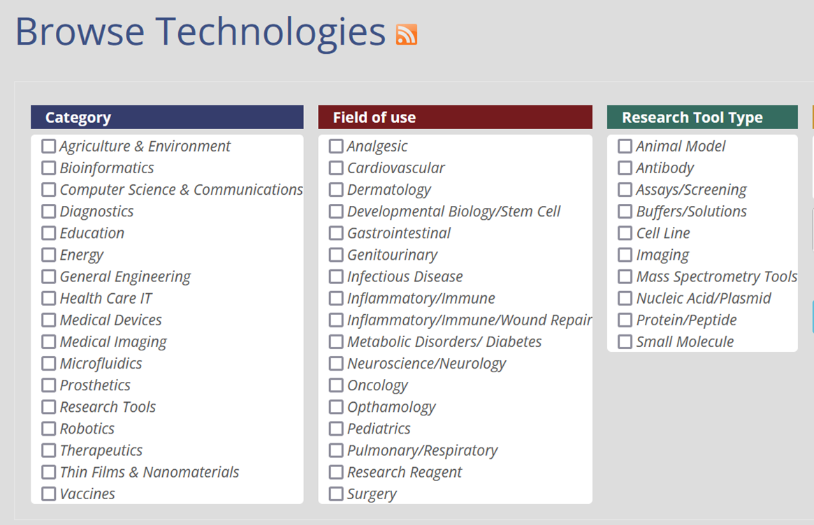
Browse Technologies

Displaying 21 - 30 of 50


PIQASO: A rigid phantom for comprehensive end-to-end evaluation of online adaptive radiotherapy systems

There is currently no radiotherapy phantom capable of quantitatively assessing all components of an online adaptive radiotherapy (online ART) system in a comprehensive end-to-end test.Represented here is a novel, rigid phantom that can simultaneously evaluate an online ART system's image acquisition, deformable image registration, contour propagation, plan re-optimization, dose calculation, and beam delivery in a single process that is robust, quantitative, and convenient.


Licensing Contact

Masood Machingal

615.343.3548

Inexpensive Disposable Hydro-Jet Capsule Robot for Gastric Cancer Screening in Low-Income Countries

Gastric cancer is the second leading cause of cancer death worldwide. While screening programs have had a tremendous impact on reducing mortality, the majority of cases occur in low and middle-income countries (LMIC). Typically, screening for gastric and esophageal cancer is performed using a flexible endoscope; however, endoscopy resources for these settings are traditionally limited. With the development of an inexpensive, disposable system by Vanderbilt researchers, gastroscopy and colonoscopy can be facilitated in areas hampered by a lack of access to the appropriate means.


Licensing Contact

Masood Machingal

615.343.3548

A Method to Obtain Uniform Radio Frequency Fields in the Body for High Field MRI

Researchers at Vanderbilt have created a new approach to produce uniform radio frequency (RF) fields in the body during high field magnetic resonance imaging (MRI). Existing high field MRI machines create non-uniform RF fields that lead to non-uniform sensitivity in the generated images, also referred to as "hot" and "cold" spots. These local variations interfere with the tissue contrast of the images that radiologists depend upon to make accurate diagnoses. By generating uniform RF fields in the body, this technology provides the benefits of high field MRI without the non-uniform RF fields.


Licensing Contact

Chris Harris

615.343.4433
Medical Imaging

Enhanced Optical Imaging for the Treatment of Retinal Disease

Vanderbilt researchers have developed a system for enhancing the imaging capabilities of optical coherence tomography (OCT), a tool commonly used to monitor and treat patients with retinal disease. The image resolution of OCT, however, is intrinsically limited. Ideally, a contrast agent could be used to highlight specific parts of the retina within the image, but dye alone is largely ineffective because of the way OCT generates the image. Photothermal heating solves this problem by creating local zones of tissue expansion which can be distinctly detected by OCT. Photothermal-OCT is safe, effective, and enhances the imaging power of a tool widely used by opticians.


Licensing Contact

Philip Swaney

615.322.1067
Medical Imaging

Systems and Methods for Non-destructive Evaluation of Optical Material Properties and Surfaces

A team of Vanderbilt researchers has developed a novel system and method for non-destructive characterization of compound lenses. The approach uses optical coherence tomography and reflectance confocal microscopy to fully characterize lens geometry and glass materials, enabling accurate modeling of compound lenses.


Licensing Contact

Philip Swaney

615.322.1067
Medical Imaging

Bioresorbable RF Coils for Post-Surgical Monitoring by MRI

Vanderbilt researchers have developed bioresorbable RF coils to improve the signal-to-noise ratio (SNR) for use in post-surgical monitoring.


Licensing Contact

Chris Harris

615.343.4433

Inventors

Mark Does, John Rogers

Innovative Mobile App that Facilitates Self-Management in Diabetes

Vanderbilt researchers have developed the MyDay mobile app (iOS/Android) designed to collect, integrate, and provide feedback on a wide range of individual data relevant for diabetes self-management which allows flexible creation of data collection content, format, and timing.


Licensing Contact

Masood Machingal

615.343.3548

Molecular Image Fusion: Cross-Modality Modeling and Prediction Software for Molecular Imaging

A research team at Vanderbilt University Mass Spectrometry Research Center has developed the Molecular Image Fusion software system, that by fusing spatial correspondence between histology and imaging mass spectrometry (IMS) measurements and cross-modality modeling, can predict ion distributions in tissue at spatial resolutions that exceed their acquisition resolution. The prediction resolution can even exceed the highest spatial resolution at which IMS can be physically measured. This software has been successfully tested on different IMS datasets and can be extended to other imaging modalities like MRI, PET, CT, profilometry, ion mobility spectroscopy, and different forms of microscopy.


Licensing Contact

Karen Rufus

615.322.4295

Self-Decoupled RF Coils for Optimized Magnetic Resonance Imaging

Magnetic resonance imaging (MRI) is one of the most important and versatile tools in the repertoire of diagnostics and medical imaging. Vanderbilt researchers have developed a novel, geometry independent, self-decoupling radiofrequency (RF) coil design that will allow MRI machines to generate images at a faster rate and with greater image quality.


Licensing Contact

Philip Swaney

615.322.1067

Advanced Method for Data Corrections in Organ Deformation

A group of Vanderbilt University researchers have developed a solution that will correct for the mis-registration of image data in image-guided surgery. The solution uses software to correct for any mis-registration that is caused by the presence of intraoperative deformations. This invention helps to improve the performance and capabilities of image-guided surgery.


Licensing Contact

Philip Swaney

615.322.1067
Medical Imaging